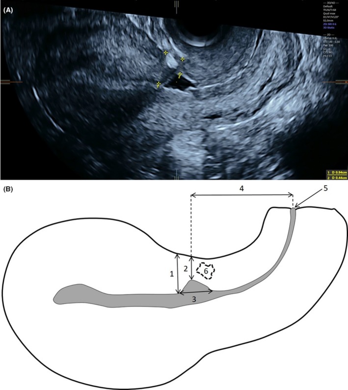
Figure 2.

(A) Transvaginal ultrasound demonstrating measurement of total myometrial thickness (1) and residual myometrial thickness (2). (B) Schematic diagram showing CS scar placement and dimensions measurement: total myometrial thickness (1), residual myometrial thickness (2), width of the scar defect (3), distance between the scar and the external cervical ostium (4), external cervical ostium (5), myometrial defects without contact with the uterine cavity (6) [Color figure can be viewed at http://wileyonlinelibrary.com]
