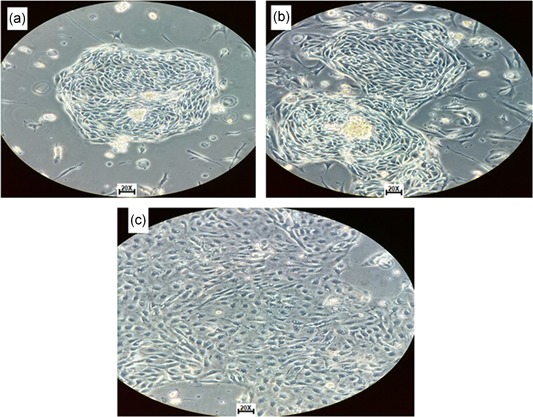
Figure 2.

Isolated cells from solid tumor morphology. (a) Cells with epithelial phenotype and raceme‐shaped growth at 6 days after the start of the culture (code number seven). (b) Culture with confluence between 70% and 90% in the form of clusters at 10 days. (c) Cells with confluence between 80% and 95% at 10 days with cobblestone shapes, typical morphology of epithelial tissue of ovary with enzymatic disaggregation with Dispase II (code number eight) [Color figure can be viewed at wileyonlinelibrary.com]
