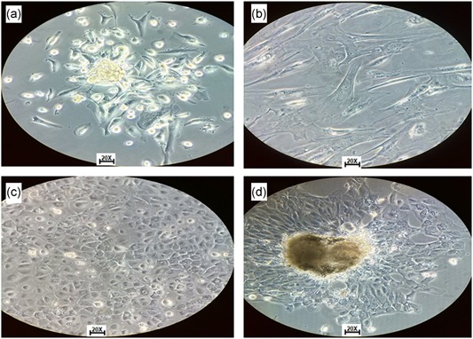
Figure 3.

Tumor cells isolated from ascitic fluid and solid tumor. (a) Isolated cells from ascitic fluid with spindle‐shaped growth. (b) Isolated cells from ascites fluid, where cellular variability is observed. (c) Isolated cells from solid tumor in DMEM/F12 medium supplemented with SFB 10%. (d) Isolated cells from ascites liquid in DMEM/F12 medium supplemented with SFB 10%. All photos were taken with inverted microscope objective ×20. DMEM, Dulbecco's modified Eagle medium [Color figure can be viewed at wileyonlinelibrary.com]
