
Figure 1.
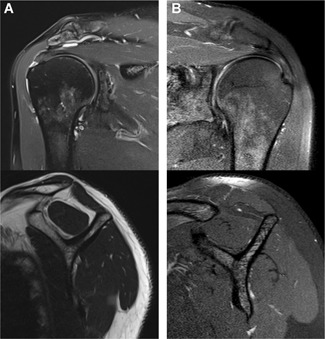
Magnetic resonance imaging of the affected shoulder. The oblique coronal imaging is shown on the top row and the sagittal images on the bottom row, respectively. (A) A full‐thickness rotator cuff tear with slight tendon retraction and mild muscle degeneration. (B) A chronic tendinopathy without any tearing of the supraspinatus tendon and muscle degeneration.