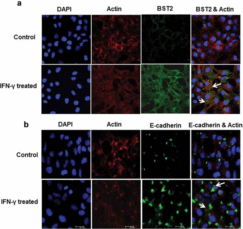
Figure 2.

Immunofluorescent profile of BST2 and E-cadherin in HTR-8/SVneo cells treated with IFN-γ. HTR-8/SVneo cells (20,000/well) were seeded on the coverslips in 24-well cell culture plates and cultured overnight at 37°C in 5% CO2 and 70% relative humidity. The next day, cells were treated with and without IFN-γ for 24 h followed by immunolocalization of BST2, E-cadherin, and actin as described in Materials and Methods. Panel a shows the expression of actin (red) and BST2 (green) in control and IFN-γ-treated HTR-8/SVneo cells. Panel b shows the expression of actin (red) and E-cadherin (green) in control and IFN-γ-treated cells. The nuclei were stained using Hoechst nuclear staining dye. Scale bar shows 20 μm.
