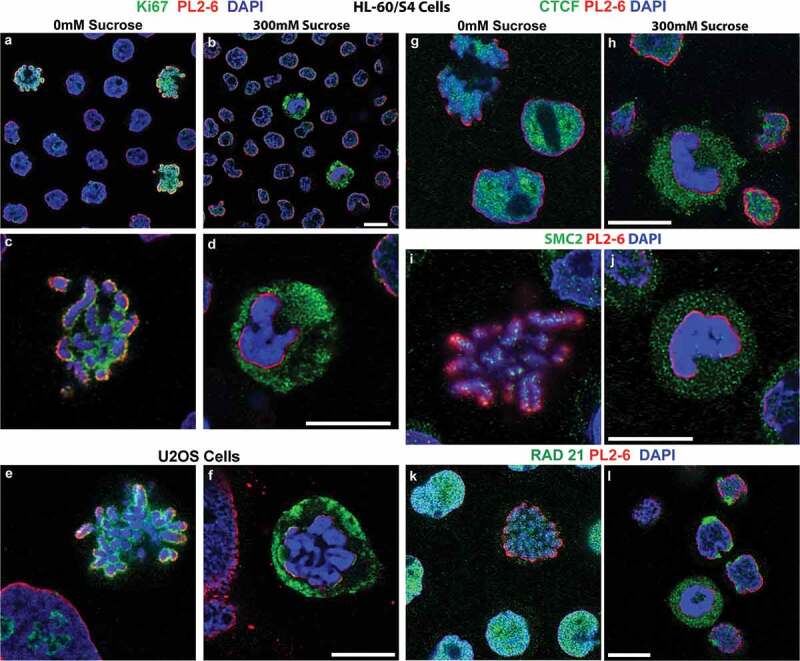
Figure 5.

Select nuclear proteins separate from hyperosmotic sucrose congealed mitotic chromatin. Rabbit antibodies against Ki67, CTCF, SMC2 and RAD21 (all green), and counterstained with PL2-6 (red) and DAPI (blue). All panels, except (e,f), show images of HL-60/S4. (a-d) Stained with anti-Ki67. (a,b), HL-60/S4 low magnification, mixed interphase and mitotic cells; (c,d), high magnification, mitotic chromosomes. (e,f) U2OS cells stained with Ki67. (a,c,e,g,i,k), 0 mM sucrose; (b,d,f,h,j,l), 300 mM sucrose. Note the Ki67 staining surrounding nucleoli of 0 mM sucrose interphase nuclei, which are absent from 300 mM sucrose interphase nuclei. (g,h), anti-CTCF. (i,j), anti-SMC2. (k,l), anti-RAD21. The upper left corner of (g) contains a cluster of mitotic chromosomes stained with anti-CTCF. Magnification bars, 10 µm.
