
Figure 7.
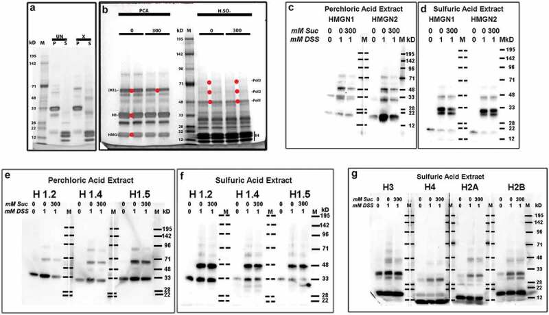
DSS cross-linking of acid extractable chromatin proteins. (a), Coomassie blue (InstantBlue) stained 4-12% acrylamide gradient SDS-PAGE of PCA (p) and H2SO4 (s) extracts from HL-60/S4 cells crosslinked (or not) with 1 mM DSS in PBS ± 300 mM sucrose. Comparison of uncrosslinked (UN) or crosslinked (x) extracts. Molecular weight markers (m) are aligned with their assigned weights (kD). Note the presumptive H1 monomer (MW ~33 kD) and the presumptive H1 dimers following DSS crosslinking (MW ~68 kD). (b), Comparison of acid extracts from HL-60/S4 cells crosslinked with 1 mM DSS in PBS (pH 8) ± 300 mM sucrose. Each sample was run in duplicate. The red dots placed between duplicate bands indicate the adjacent gel regions that were excised for mass spectroscopy: for 0 mM sucrose [HMG, H1 and (H1)2]; for 300 mM sucrose [(H1)2, Pol1, Pol2 and Pol3]. ‘Pol’ bands represent presumptive ‘polymers’ of the histones and/or HMG proteins (see Supplementary Table S1). IH, inner histone (H3, H4, H2A, H2B) region. Note that, for panel (b), the DSS crosslinked products show considerable similarity, ± exposure to hyperosmotic sucrose. (c,d) Immunoblots of anti-HMGN1 and anti-HMGN2 reacted with PVDF membranes from 4-12% SDS-PAGE of PCA (c) or H2SO4 (d) extracts of HL-60/S4 cells crosslinked (or not) with 1 mM DSS in PBS ± 300 mM sucrose. Indicated at the top of each lane are the mM sucrose and the mM DSS employed in that preparation. Note the presumptive HMG monomers (MW ~20 kD) and the presumptive DSS crosslinked products (MW ~33 and ~48 kD) within the PCA (c) and H2SO4 (d) extracts. Also note that, for panels (c) and (d), these DSS crosslinked products show considerable similarity, ± exposure to hyperosmotic sucrose. Crosslinked products do not appear in the uncrosslinked extracts, except for HMGN1, which showed a weak band at ~33 kD in the PCA extract. (e,f) Immunoblots of anti-H1 antibodies reacted with PCA (e) or H2SO4 (f) extracts of HL-60/S4 cells. Note that for each histone H1, the DSS crosslinked products show considerable similarity, ± exposure to hyperosmotic sucrose. (g) Immunoblots of anti-inner histone (H3, H4, H2A and H2B) antibodies reacted with H2SO4 extracts of HL-60/S4 cells. Note that for each inner histone, the DSS crosslinked products show considerable similarity, ± exposure to hyperosmotic sucrose.