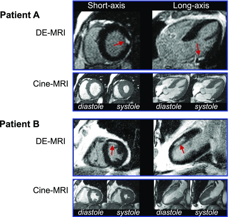
Figure 1.
Typical DE-MRI and cine-MRI images. Short- and long-axis views in two patients are shown. Patient A demonstrates a small, focal MI (arrows) limited to the basal anterolateral wall. Patient B has a subendocardial infarction (arrows) involving the mid-anterior wall. Both patients had normal LVEF (A, 64%; B, 61%), no regional wall motion abnormalities, and no Q waves on electrocardiography.

