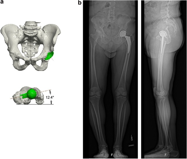
Fig. 1.
a 3D computed tomography (CT) reconstruction of pelvis (coronal view) and femur (transverse view). Implants are shown in green; CT femoral anteversion in this patient was 12.4°. b Simultaneous and orthogonal acquisition of these standing anteroposterior and lateral radiographs allowed for 3D implant position measurements (cup inclination, cup anteversion, and femoral anteversion).

