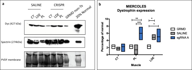
Fig 5. Dystrophin protein expression after HDR-CRISPR treatment in Miercoles.
CRISPR sgRNA A was injected into one cranial tibial compartment and saline into the other. The vastus lateralis muscle from Miercoles (GRMD) was biopsied before injections to provide a general baseline and the HDR-CRISPR/saline muscles were harvested 3 months after treatment. (a) Dystrophin co-stained with C and N-terminus antibodies with goat anti-mouse secondary staining, β-spectrin stained as a muscle marker with goat anti rabbit secondary staining. Total protein from the PVDF membrane was used to normalize dystrophin. Normal sample was diluted to 20%. (b) Graph with dystrophin quantification for each muscle in the cranial tibial compartment. The PL and LDE muscles had increased dystrophin restoration in the HDR-CRISPR-Tx muscle compared to saline and GRMD while there were no differences in the HDR-CRISPR-Tx CT muscle. Statistical analysis was performed with Tukey’s multiple comparison’s test * p ≤ 0.05;** p ≤ 0.01;*** p ≤ 0.001. CT = cranial tibial; LDE = long digital extensor; PL = peroneus longus.

