. 2020 Jan 21;9:e51701. doi: 10.7554/eLife.51701

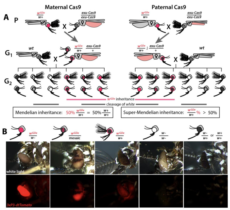
Figure 2. Assessing super-Mendelian inheritance of split drives.

(A) Crossing scheme of the wGDe and exu-Cas9 parent strains (P) to generate trans-heterozygotes (G1), and the outcrossing of their progeny to wild-type (wt) mosquitoes (G2). tdTomato eye and dsRed abdominal expression were the wGDe and exu-Cas9 transgene inheritance markers, respectively. wGDe transmission and white loss-of-function mutation rates were estimated among G2 progeny of trans-heterozygous female and wt male crosses. Super-Mendelian inheritance of wGDe occurred when the transmission rate of wGDe in G2 progeny was >50%, as expected by standard Mendelian inheritance. (B) Examples of G2 progeny eye phenotypes and corresponding genotypes.

Figure 2.

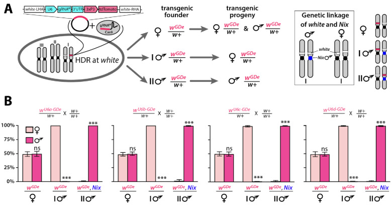
Figure 2—figure supplement 1. Sex biased inheritance of the GDe is due to linkage of white and Nix, a male determining gene in Ae. aegypti.

Figure 2—figure supplement 1.

Schematic of site-specific integration and sex-linked inheritance of Gene Drive element (GDe) (A). To direct site-specific integration of GDe inside white locus (white stripe on chromosome I) via homology directed repair (HDR), wild type (wt) Ae. aegypti embryos were injected with a Cas9/gRNAw complex and a plasmid carrying GDe flanked by homology regions complementary to left and right genomic regions at the white cut site. GDe contains two genes: U6-gRNAw to direct a site-specific cleavage and the 3xP3-tdTomato transgenesis marker (red stripe). Female mosquitoes carrying wGDe/w+ passed wGDe randomly to both genders, while transgenic males transmitted wGDe either to females (type I) or males (type II) nearly exclusively. Close genetic linkage of white and Nix genes causes tight sex-linked inheritance of wGDe via male founders (B). Nix is located near the centromere of chromosome I (Dudchenko et al., 2017; Matthews et al., 2018) and consequently, type I male () founders have wGDe inserted on the chromosome copy without Nix and therefore pass wGDe exclusively to female progeny. On the other hand, type II male founders have wGDe integrated on the chromosome with Nix, and thus they transfer wGDe and Nix together exclusively to male progeny (box). Bars show average ± SD estimated for 20 data points. Statistical significance between gender frequencies was estimated by an equal variance t test. (P ≥ 0.05ns and p<0.001***).
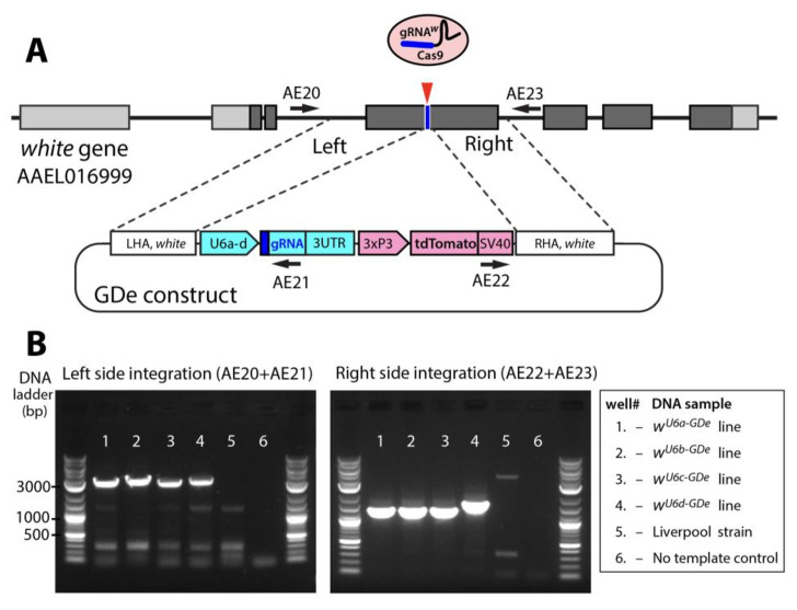
Figure 2—figure supplement 2. Site-specific integration of Gene Drive element (GDe).

Figure 2—figure supplement 2.

Schematic showing positions of Left and Right Homology Arms (LHA and RHA) of GDe relative to genomic white sequence. The cut site is presented over the genetic structure of white, with darker gray boxes representing exonic coding sequences. GDe constructs were integrated at the cut site via homology directed repair (HDR) relying on the construct’s LHA and RHA to white. Arrows depict primers (AE20, AE21, AE22 and AE23) used for PCR and their location relative to genomic and constructs sequences. Two gel images showing specific PCR amplicons for each side of integration (B). The same DNA samples, listed in the box to the left, were used to PCR of both fragments. To confirm PCR specificity, amplicons were Sanger sequenced from both ends.