Figure 1.
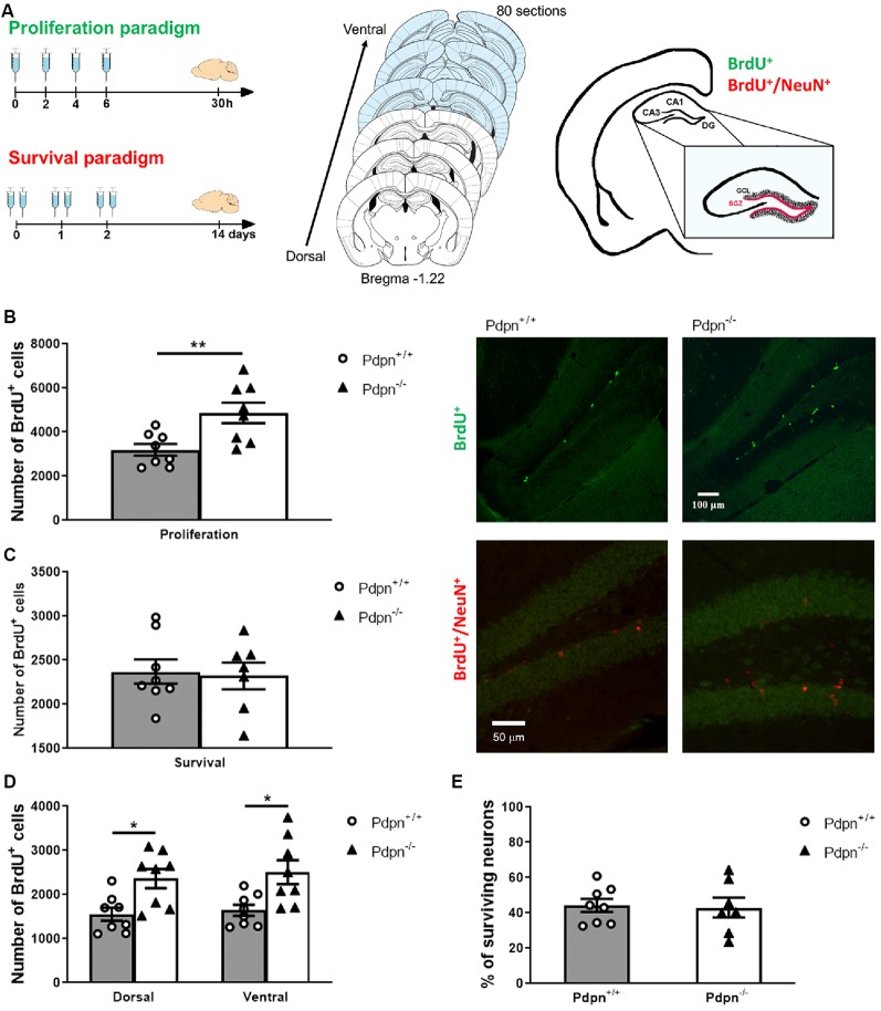
Lack of Podoplanin increases the proliferation of cells in the subgranular zone (SZG) of the dentate gyrus (DG). (A) Schematic representation of 5-bromodeoxyuridine (BrdU) injection protocols for proliferation and survival paradigms (left panel). Schematic representation span of the rostrocaudal axes from which coronal sections were collected (middle). The depicted coronal sections have been modified from Franklin and Paxinos (2013). The SGZ spans at the border of the granule cell layer (GCL) and hilus of the hippocampal DG (right panel). (B) Analysis of the approximated number of BrdU+ cells per hippocampus in “proliferation” paradigm showed a significant difference between two genotypes (**P = 0.008, t(14) = 3.122, n = 8 per group). Representative photomicrographs of Pdpn+/+ and Pdpn−/− coronal sections immunostained against BrdU in the “proliferation” paradigm (right panel 10× magnification). (C) In the “survival” paradigm quantification of BrdU+ cells showed no differences between hippocampi of Pdpn+/+ and Pdpn−/− mice (P = 0.819, t(13) = 0.233, n = 7–8 per group). Representative photomicrographs of Pdpn+/+ and Pdpn−/− coronal sections immunostained against BrdU in red and against NeuN in green in the “survival” paradigm (right panel 20× magnification). (D) Two-way ANOVA of the number of BrdU+ cells in “proliferation” paradigm between dorsal and ventral hippocampus showed a main significant effect of genotype (***P < 0.0002, F(1,28) = 17.61, n = 8 per group), no significant effect of the area (P = 0.5601, F(1,28) = 0.3477, n = 8 per group) and no significant interaction between paradigm and genotype (P = 0.8912, F(1,28) = 0.0190, n = 8 per group). Post hoc analysis showed significant difference between Dorsal:Pdpn+/+ and Dorsal:Pdpn−/− (*P = 0.0366), Dorsal:Pdpn+/+ and Ventral:Pdpn−/− (*P = 0.0108), as well as Ventral:Pdpn+/+ and Ventral:Pdpn−/− (*P = 0.0233), as represented with asterisks on the graph. (E) Quantification of surviving cells double-labeled with BrdU+ and NeuN+ showed no significant difference in a number of surviving neurons in Pdpn−/− mice compared to their Pdpn+/+ littermates (t(13) = 0.1857, P = 0.9, n = 7–8). Data are displayed as mean ± SEM.

