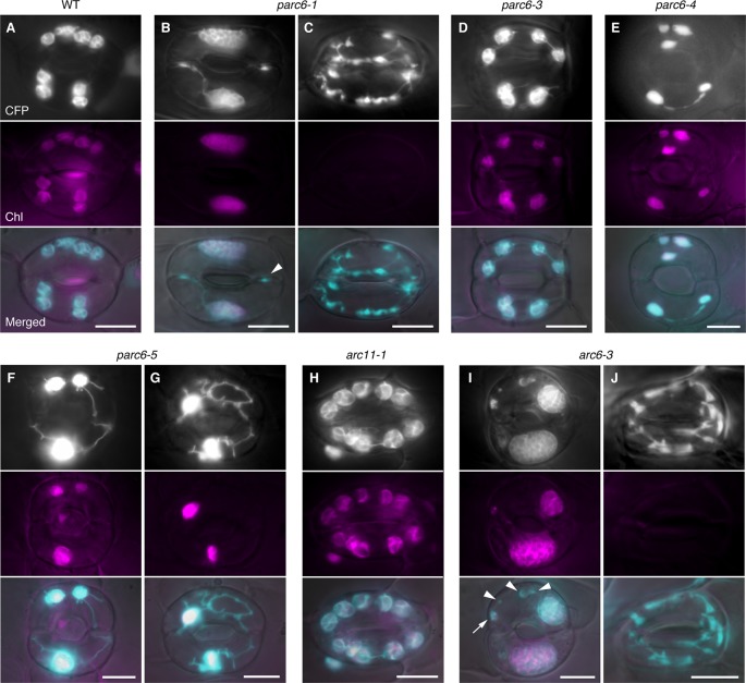
Figure 4.
Morphology of plastids in leaf stomatal guard cells of parc6 and other plastid division mutants. (A–J) Images of guard cells in the 3rd and 4th leaf petioles of 4-week-old WT (A), parc6-1 (B, C), parc6-3 (D), parc6-4 (E), parc6-5 (F, G), arc11-1 (H), and arc6-3 (I, J) seedlings. Fluorescence images of stroma-targeted CFP (black-and-white or cyan-colored in ‘merged’ panels), chlorophyll (magenta), and merged images of CFP/YFP, chlorophyll and DIC are shown. Arrow and arrowheads indicate plastids with and without chlorophyll, respectively. Scale bar = 10 µm.

