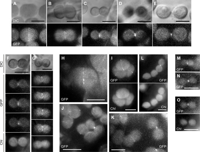
Figure 7.
Analysis of PARC6-GFP localization in leaf cortex and pavement cells. (A–O) Images of chloroplasts in leaf petioles of 1–3-week-old seedlings of parc6-1 complementation lines. Images of full-length PARC6-GFP, chlorophyll, or DIC in cortex (A–K) and pavement cells (M–O) are shown. Scale bars: 10 µm (J); 5 µm (others).

