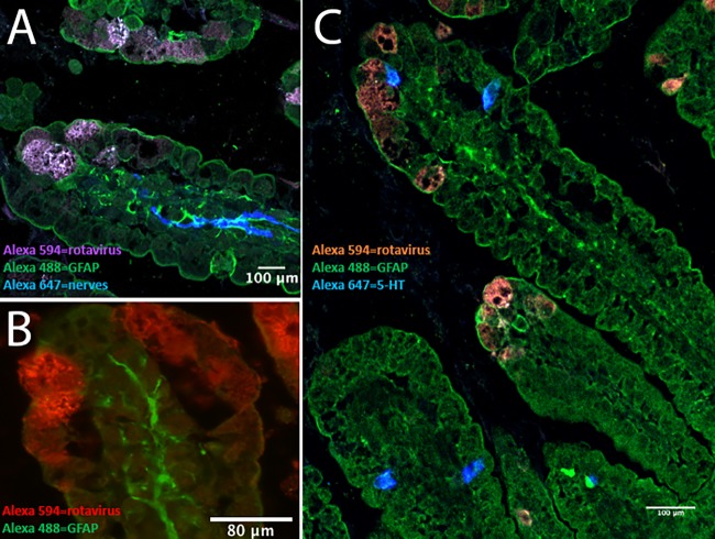
FIG 2.
EGCs and nerves are in close proximity to EC cells and rotavirus-infected enterocytes in mouse small intestine. Infant BALB/c mice (5 to 7 days old) were infected for 24 h with 100 DD of murine rotavirus (strain EDIM), and the small intestine was processed for immunofluorescence. (A) Rotavirus-infected enterocytes (purple) in the duodenum are in close proximity to activated EGCs (green, GFAP staining), and activated EGCs are in proximity to the enteric nerves (blue). (B) Rotavirus-infected enterocytes (red) in the duodenum and activated EGCs (green, GFAP staining). (C) Rotavirus-infected cells (red), EC cells (blue, 5-HT staining), and activated EGCs (green, GFAP staining).

