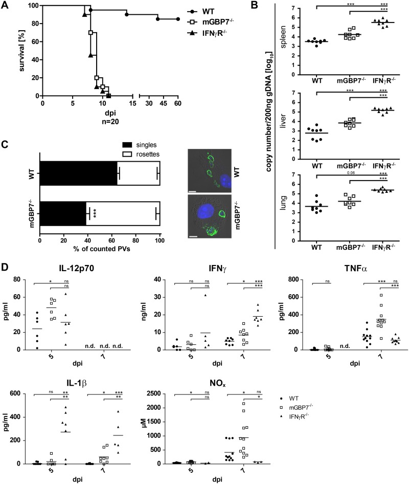
FIG 1.
Analysis of mGBP7−/− mice after T. gondii infection. (A) WT, IFN-γR−/−, and mGBP7−/− mice were infected i.p. with T. gondii (40 cysts of strain ME49) and monitored for 60 days. The survival rate of WT, IFN-γR−/−, and mGBP7−/− mice is shown (n = 20 per group). P < 0.0001. A combination of two independent experiments is depicted. (B) qPCR of T. gondii B1 gene copy numbers per 200 ng DNA from splenic, lung, and liver tissue of infected WT, mGBP7−/−, and IFN-γR−/− mice at day 7 after i.p. infection with 40 T. gondii (ME49) cysts. Specific primers and probe, and a B1 gene plasmid as an external standard, were used (n = 3). Log-transformed data are shown. The mean is marked with a bar. (C) WT and mGBP7−/− BMDMs were pretreated overnight with IFN-γ and infected with T. gondii ME49 for 24 h. T. gondii was detected with α-SAG1 monoclonal antibody (MAb), and nuclei were stained with DAPI. The percentage of intracellular T. gondii rosettes and the percentage of PVs containing a single parasite were enumerated microscopically from at least 14 field views per genotype. Insets show representative examples of infected WT or mGBP7−/− BMDMs. Means ± SEM are shown. (D) WT, IFN-γR−/−, and mGBP7−/− mice were infected i.p. with T. gondii (40 cysts of strain ME49). At the indicated time points, sera of the mice were collected, and the cytokine levels of IL-12p70, IFN-γ, TNF-α, and IL-1β were measured by ELISA. Additionally, NOx levels were measured by photometric detection (Griess reagent). At least 5 mice per group were tested. The mean is marked with a bar. *, P ≤ 0.05; **, P < 0.01; ***, P ≤ 0.001; ns, not significant; n.d., not detectable.

