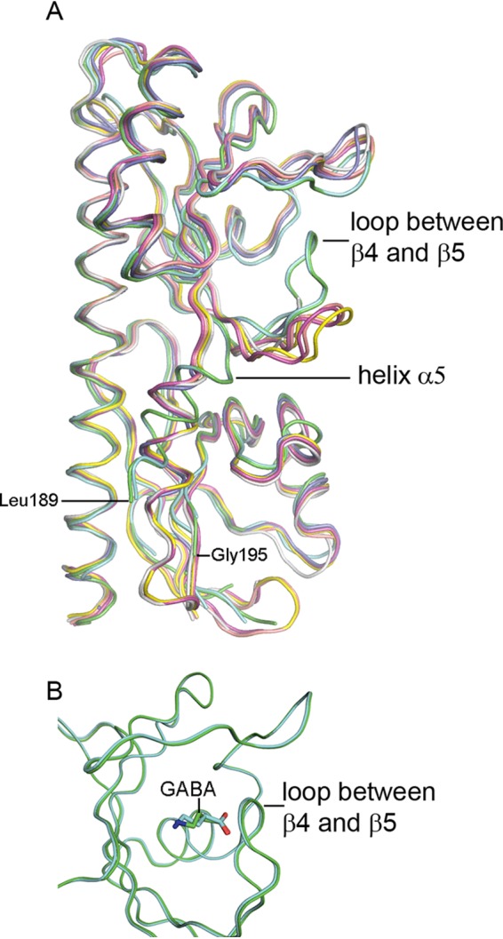
FIG 4.

Evidence for ligand-induced structural changes in PctC-LBD. (A) Structural alignment of the seven PctC-LBD chains of the asymmetric unit. Green and cyan, GABA inside the binding pocket; pink, yellow, and salmon, GABA at the entrance of the binding pocket; gray, acetate in the binding pocket; blue, empty pocket. (B) Superimposition of the membrane-distal modules of the two PctC-LBD chains with GABA inside the binding pocket.
