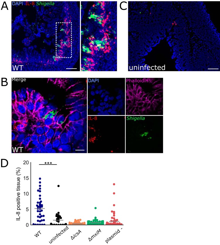
FIG 4.
Colonic IL-8 mRNA in rabbits infected with S. flexneri. (A to C) Immunofluorescence micrographs of colonic sections from infant rabbits infected with WT S. flexneri (A and B) or uninfected control (C). Sections were stained with an RNAscope probe to rabbit IL-8 (red), an antibody to Shigella (FITC-conjugated anti-Shigella green), and with DAPI (blue). (A) Colon section infected with WT S. flexneri. The inset to the right in panel A depicts a magnified view of the boxed area in the left image. Bar, 200 μm. (B) High magnification of colonic epithelium infected with WT S. flexneri. Sections were also stained with anti-E-cadherin antibody (magenta). Bar, 10 μm. (C) Uninfected colon section. Bar, 100 μm. (D) Percentage of IL-8-expressing cells in each field of view from colonic tissue sections stained with probe to rabbit IL-8 from rabbits infected with the indicated strain. See Materials and Methods for additional information regarding the determination of these measurements. Mean values are indicated with bars. All groups were compared to the sections from the uninfected animals. Statistical significance was determined using a Kruskal-Wallis test with Dunn’s multiple-comparison posttest. ***, P < 0.001.

