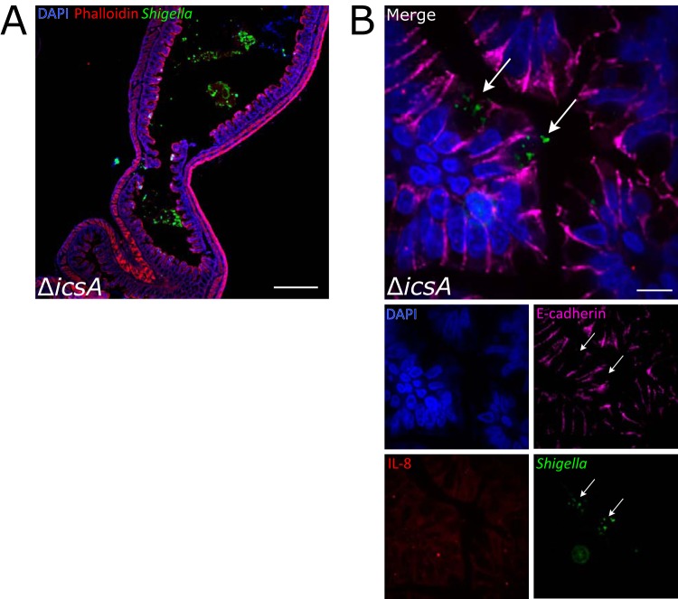
FIG 7.
Intestinal localization and IL-8 transcripts in colons from animals infected with an icsA mutant. (A) Immunofluorescence micrograph of ΔicsA in colonic tissue of infected rabbits 36 hpi. DAPI (blue), FITC-conjugated anti-Shigella antibody (green), and phalloidin-Alexa Fluor 568 (red) are shown. Bar, 500 μm. (B) Immunofluorescence micrograph of sections stained with a RNAscope probe to rabbit IL-8 (red) and antibodies to Shigella (green) and E-cadherin (magenta), and DAPI (blue). The bottom panels depict channels of the merged image. Arrows point to multiple icsA bacteria in the cytoplasm of two infected cells. Bar, 10 μm.

