FIGURE 4.
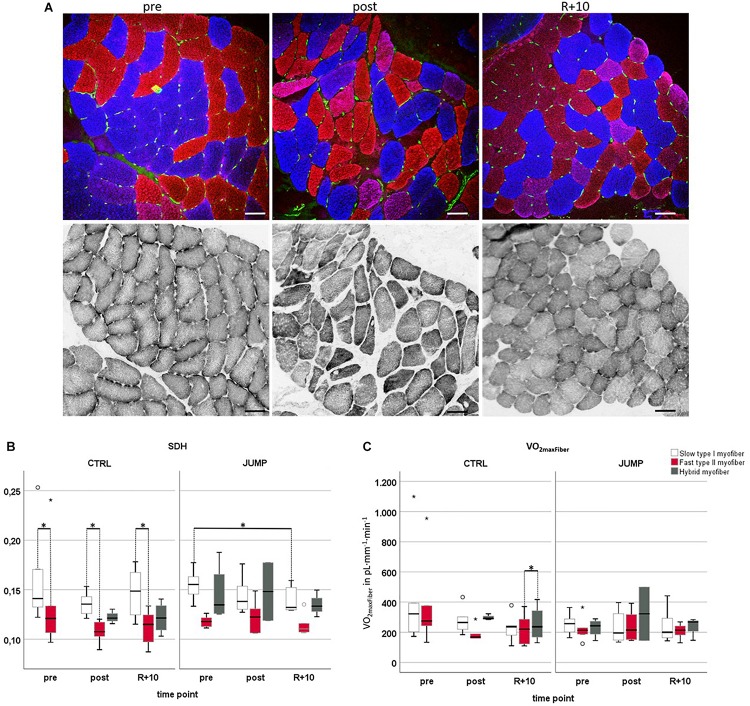
Oxidative capacity analyzed by semiquantitative histochemical succinate dehydrogenase optical density analysis (ODSDH) using RSL study biopsy cryosections. (A) Representative subject-matched/paired microscopic images at three different time points [vastus lateralis (VL), pre/post/R + 10]. Upper panel, immunostaining for slow MyHC-I (red), fast MyHC-II (blue), and capillary platelet endothelial cell adhesion molecule 1 (PECAM-1; green). Lower panel, adjacent cryosections with matched area stained for SDH histochemical activity at 37°C, index of mitochondrial activity (gray values). Scale bars = 75 μm. (B) Determination of optical density of SDH marker at 660 nm (OD660) in control (CTRL) (myofiber type I/II n = 6, hybrid pre n = 0 post n = 3 R + 10 n = 4) or exercise (JUMP, VL myofiber type I/II n = 6, hybrid pre n = 4, post n = 2, R + 10 = 3). The specific SDH activity (ODSDH) was always higher in CTRL pre/post/recovery (R + 10) type I vs. type II myofibers (pre p = 0.028, post p = 0.043, recovery p = 0.014). In the JUMP group, the amount of SDH activity was significantly different (reduced) in slow myofibers at R + 10 vs. pre (p = 0.028). (C) VO2maxFiber values showed a significant difference between recovery fast < hybrid (p = 0.014) with no changes in JUMP. CTRL group (left, myofiber type I/II n = 6, hybrid pre n = 0, post n = 3, R + 10 n = 4), JUMP group (right, myofiber type I/II n = 6, hybrid pre n = 4, post n = 2, R + 10 = 3). SDH and VO2maxFiber of slow type 1 (red), fast type 2 (blue), and hybrid myofibers (green) in participants without (CTRL) and with exercise (JUMP) at pre/post/recovery time points of head-down tilt (HDT) bed rest. ∗Significance at p < 0.05, box plots (means) with median ± 2 SE.

