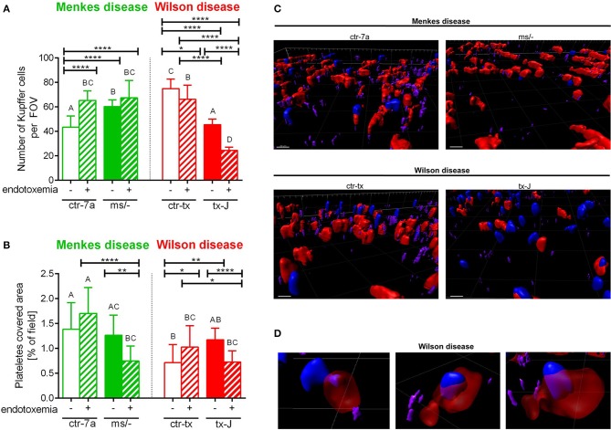
Figure 4.
The presence of Kupffer cells (A) and platelets (B) in livers of Wilson (tx-J) and Menkes (ms/−) disease mice and their respective controls (ctr-tx and ctr-7a) with endotoxemia. Kupffer cells were counted per field of view (FOV), and area covered by platelets is expressed in percentage (as calculated by ImageJ) (mice n = 3/group). Asterisks indicate significant differences using unpaired two-tailed Student's t test (*P ≤ 0.05, **P ≤ 0.01, ****P ≤ 0.0001) and one-way ANOVA (post hoc Bonferroni) test (different letters indicate statistically significant differences between groups). A series of optical cross-sections (z stacks) were made through inflamed livers. Exemplary images of z stacks made through the liver of Menkes disease and Wilson disease mice and their respective controls are shown in (C). Kupffer cells are marked in red, neutrophils in blue, and neutrophil elastase in violet. In endotoxemic mice with Wilson disease, neutrophils present (partially or fully) inside some Kupffer cells were observed (D). The scale bar indicates 20 μm.

