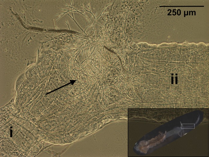
FIG 1.
Zancudomyces culisetae colonization of a dissected fourth-instar larval mosquito digestive tract (magnified view of area boxed in inset image, bottom right) visualized with phase-contrast microscopy (×100 magnification). Significant regions are labeled as follows: i, midgut; ii, hindgut. The black arrow indicates mature fungal hyphae. The inset shows a larva with the head removed and midgut and hindgut exposed.

