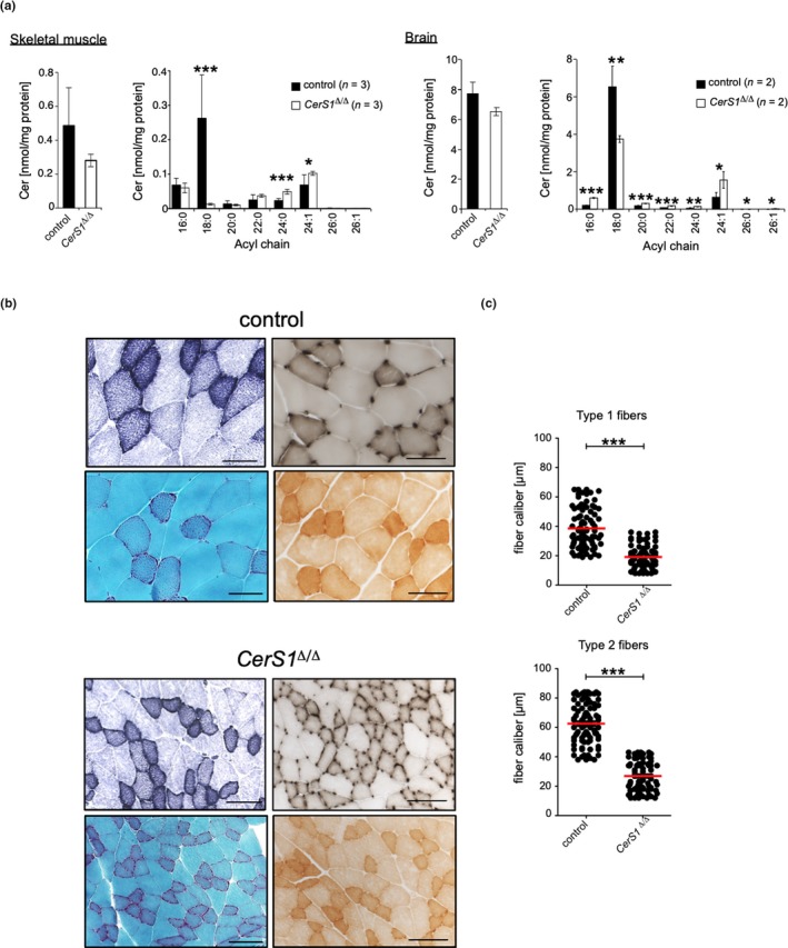
Figure 3.

CerS1 deficiency leads to prominent C18:0 Cer depletion in skeletal muscle and brain and is associated with muscle atrophy. (a) Quantitative analysis of changes in acyl chain length distribution of Cer in skeletal muscle or brain of male CerS1 Δ/Δ mice, represented is mean ± SD, exact number of mice per genotype 1s indicated in figure. Each tissue sample was analyzed in duplicates. Histological analysis and determination of fiber caliber size of the quadriceps femoris muscle from CerS1 Δ/Δ (n = 3m) and wild‐type control mice (n = 3m) in the age of 47 and 52 weeks old. (b) Sections were stained with red. NADH histochemistry (upper left), ATPase pH 4.4 (upper right) Gomori trichrome (lower left), or COX (lower right). Micrographs: original magnification ×200, scale bar corresponds to 100 µm. (c) Graphs depict fiber caliber size for type 1 (top) and type 2 fibers (bottom). To determine the caliber of muscle fibers (µm), serial sections with 25 high‐powered fields (HPF) of the quadriceps muscle with at least 80 muscle fibers of type 1 and type 2 fibers were counted. Each dot indicates the caliber of individual fibers, and the red bar corresponds to the mean. Statistical significance was assessed by two‐tailed unpaired Student's t test (*p < .05; **p < .01; ***p < .001)
