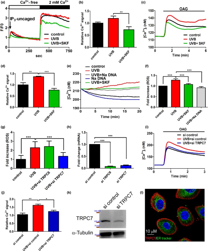
Figure 2.

Ultraviolet B (UVB)‐induced [Ca2+] elevation in keratinocytes mediated specifically by Ca2+ influx via TRPC7. (a) UVB exposure had no effect on uncaged inositol trisphosphate (IP3)‐induced activation of TRPC1, TRPC4, and TRPC5 or the subsequent [Ca2+]i elevation in keratinocytes. SKF96365 pretreatment (positive control) significantly decreased UVB‐induced [Ca2+]i elevation. (b) The mean (±SD) area under the [Ca2+]i response curves from 60 cells after the addition of extracellular CaCl2. (c) In contrast, UVB pre‐exposure significantly increased [Ca2+]i elevation in response to 1‐oleoyl‐2‐acetyl‐sn‐glycerol (OAG), an effect that was significantly inhibited by SKF pretreatment. (d) The mean (± SD) area under the [Ca2+]i response curves from 235 cells after the addition of OAG. The knockdown of TRPC7 significantly decreased UVB‐induced cell damage via the reduction in the p53 pathway activation. UVB‐induced Ca2+ elevation via TRPC7 channels initiated intracellular reactive oxygen species (ROS) production in keratinocytes. (e) The time course of the increased Ca2+ response after UVB exposure demonstrated an immediate UVB‐induced increase in the intracellular Ca2+ concentration that was inhibited by pretreatment with the TRPC7 inhibitors SKF96365 (SKF) or Na DNA. (f) Intracellular ROS production began after 30 min of UVB exposure. Cells were stained with 5 μM dihydroethidium (free radical indicator), and the intensity of emitted fluorescence was analyzed by using an Olympus fluorescence microscope (n > 200 cells; mean ± SD). (g) Intracellular ROS production increased after UVB irradiation (n > 200 cells; mean ± SD). This effect was significantly reduced by the knockdown of TRPC7, but not the knockdown of TRPC6. (h) The knockdown of TRPC6 and TRPC7 mRNA expression was effective. (i) TRPC7 knockdown attenuated the OAG‐induced increase in [Ca2+]i after UVB exposure. (j) The mean area under the [Ca2+]i response curves from 260 cells after OAG application. (k) Western blot analysis showing that protein expression was reduced after TRPC7 knockdown in keratinocytes. (l) Immunofluorescence staining showing the localization of TRPC7 (red) to the cell membrane of keratinocytes. The endoplasmic reticulum is green (ER‐Tracker; 1,3,5,7‐tetramethyl‐8‐phenyl‐4,4‐difluoroboradiazaindacene; BODIPY), and nuclei are blue (4′,6‐diamidino‐2‐phenylindole; DAPI). si, small interfering RNAs. *p < .05; **p < .01; ***p < .001
