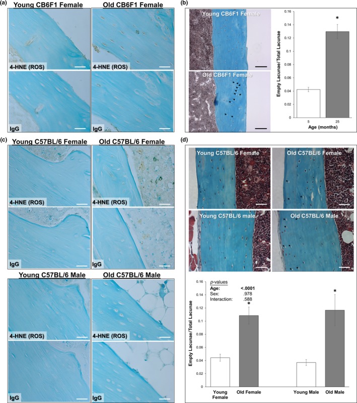
Figure 4.

Aged osteocytes are exposed to greater oxidative stress in vivo. Longitudinal tibial sections of old and young mice used for the treadmill running experiments (a–b) and for isolation of primary osteocytes (c–d) were stained to detect 4‐HNE (brown staining, panels a and c), a marker of lipid peroxidation and oxidative stress. Representative samples from each group are shown. Little to no staining was observed with a nonspecific IgG control. Counterstain: Fast Green, 400× original magnification. Additional sections were stained with Goldner's Trichrome (panels b and d) for detection and quantification of empty osteocyte lacunae. Representative images are shown; empty osteocyte lacunae are highlighted by arrows. Scale bars: 30 µm (white), 100 µm (black). *p < .05 versus sex‐matched young control
