
Figure 5.
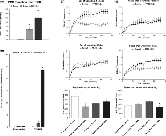
Selective survival of fast‐repairing osteocytes following turbulent fluid shear stress (TFSS) and oxidative stress. (a) MLO‐Y4 osteocytes were subjected to 5 or 20 cycles of TFSS, as described (Mikolajewicz et al., 2018), in the presence of 10 kDa fluorescein‐conjugated dextran. Control treated cells were subjected to one round of gentle media displacement in the presence of 10 kDa fluorescein‐conjugated dextran. *p < .05 versus control. (b) MLO‐Y4 osteocytes were subjected to 20 cycles of TFSS in the presence or absence of oxidative stress from hydrogen peroxide (1 mM H2O2). Cell death, as measured by comparison of quantified cell number before and after TFSS application (Panel b), was increased by the combination of TFSS + oxidative stress. *p < .05 versus nonwounded control, average of n = 3 independent experiments is shown. (c) Young female osteocytes treated with TFSS + H2O2 (1 mM) demonstrated faster rates of membrane repair as compared to control treated cells on the day of wounding, with significantly smaller area under the curve (AUC). Males, in contrast, showed no differences between groups on the day of wounding. (d) Young male osteocytes treated with TFSS + H2O2 demonstrated faster rates of membrane repair as compared to control treated cells, with significantly smaller area under the curve (AUC), when repair rate was measured three days after wounding. This trend was not seen in females. *p < .05 versus sex‐matched control group. N = 3 independent cell lines per sex