Figure 3.

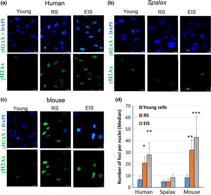
Distinct DDR in Spalax, human, and mouse fibroblasts undergoing replicative senescence (RS) and etoposide‐induced senescence (EIS). To induce senescence, young cells were treated with 1 μg/ml of etoposide for 5 days; then, etoposide was washed out and cells were replated and incubated for an additional 48 hrs. Thereafter, cells were fixed and stained with anti‐phospho‐H2AX antibody and counterstained with DAPI. Cells' nuclei were visualized under fluorescent microscope. The images were used for quantification using FociCounter software (a minimum of 250 nuclei per sample were analyzed). Representative images of γH2AX (Ser139) foci in the nuclei of young, RS, and EIS human (a), Spalax (b), and mouse (c) fibroblasts are demonstrated. (d) A quantitative analysis of foci numbers. Data are presented as median of 250 nuclei counted for each species, assessed in three independent experiments (n = 3). *** p ≤ .001, ** p ≤ .01, * p ≤ .05, differences between the groups of young and senescent nuclei. Scale bars are 25 µm
