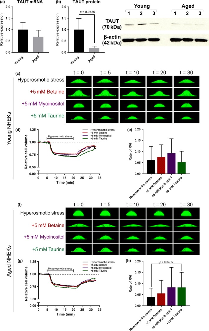
Figure 4.

TAUT protein is reduced in aged NHEKs and taurine improves the rate of cell volume recovery in aged NHEKs exposed to hyperosmotic stress. (a) Relative mRNA expression of TAUT in young and aged NHEKs. (b) Western blot for TAUT protein expression revealed a significant downregulation in aged NHEKs compared to young NHEKs (p = .0480). Single‐cell live imaging was used to assess cell volume regulation in (c‐d) young and (f‐g) aged NHEKs through a 32‐min cycle of 5 min isosmotic conditions (300mOsm per L), 15 min hyperosmotic conditions (500mOsm per L), and 12 min isosmotic conditions in the absence and presence of the organic osmolytes. (e) The rate of RVI of young NHEKs. (h) For aged NHEKs, the rate of RVI significantly increased higher in the presence of taurine (p = .0485) compared to hyperosmotic stress alone. Data expressed as mean ± SD ((a,b) Student's t test, (e, h) one‐way ANOVA) (a, b) n = 3 young and 3 aged donors, (c‐e) n = 30 cells from three young and (f‐h) n = 30 cells from three aged donors. NHEKs, normal human epidermal keratinocytes; RVI, regulatory volume increase; t, time (in minutes); TAUT, taurine transporter
