Figure 3.

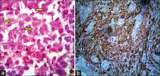
(a) The presence of sheet-like proliferation of Langerhans cells, having coffee bean-shaped appearance, eosinophils, and plasma cells (H and E, ×100). (b) Langerhans cells exhibiting positivity for anti-CD1a (×40)

(a) The presence of sheet-like proliferation of Langerhans cells, having coffee bean-shaped appearance, eosinophils, and plasma cells (H and E, ×100). (b) Langerhans cells exhibiting positivity for anti-CD1a (×40)