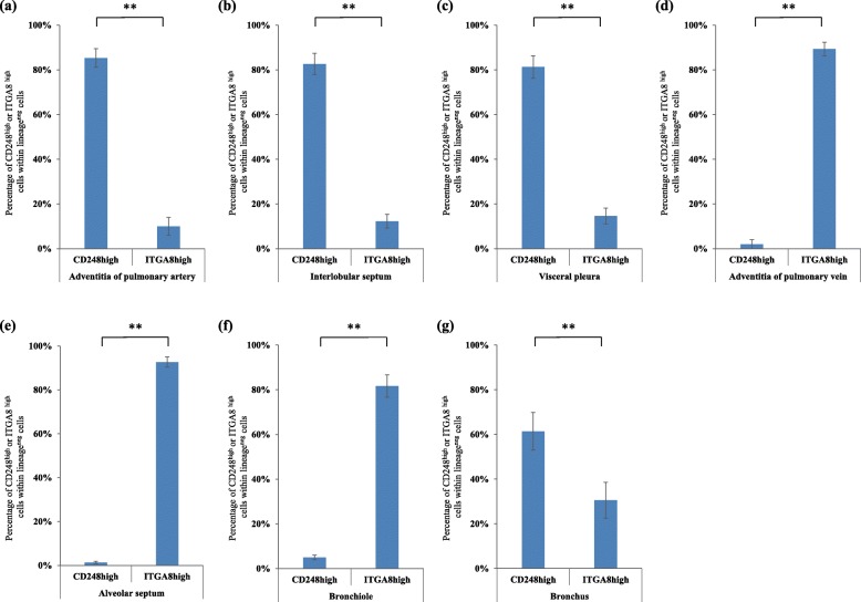
Fig. 8.
Quantification of CD248highITGA8low fibroblast-like cells and CD248lowITGA8high fibroblast-like cells in the seven regions of normal human lungs (n = 4) in the multiplex immunofluorescence staining. The ratio of CD248highITGA8low fibroblast-like cells within lineage-specific markers-negative cells (30 cells) and CD248lowITGA8high fibroblast-like cells within lineage- specific markers-negative cells (30 cells) were calculated. (a) Adventitia of pulmonary artery, (b) Interlobular septum, (c) Visceral pleura, (d) Adventitia of pulmonary vein, (e) Alveolar septum, (f) Bronchiole wall, (g) Bronchial wall. CD248high and ITGA8high (a–g) indicate CD248highITGA8low fibroblast-like cells and CD248lowITGA8high fibroblast-like cells, respectively. All experiments were performed in triplicate, and means ± standard deviations of the results obtained in three independent experiments are presented; **P < 0.01

