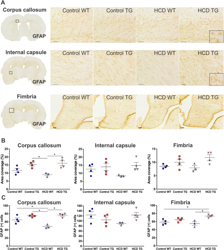
Fig. 6.
Reactive astrocytosis in white matter. a 10× photomicrographs of representative GFAP immunolabelled astrocytes in the corpus callosum, internal capsule and fimbria hippocampi. Scale bar 100 μm. Magnified images of individual astrocytes are inserted at the bottom right corner of image panels in a. b Area coverage by a positive signal (as percentage of a total area of a region) for corpus callosum, internal capsule and fimbria. Animal numbers are as follows: control WT (n = 4), control TG (n = 4), HCD WT (n = 3), HCD TG (n = 4). Values are presented as mean ± SEM. Significance is indicated by * for HCD WT vs both TG groups (in b) and additionally vs. control WT in the corpus callosum (in c); HCD TG vs both WT groups in the internal capsule (in c). One-way ANOVA and Tukey’s multiple comparisons test, p < 0.05. HCD hypercaloric diet, TG transgenic, WT wildtype

