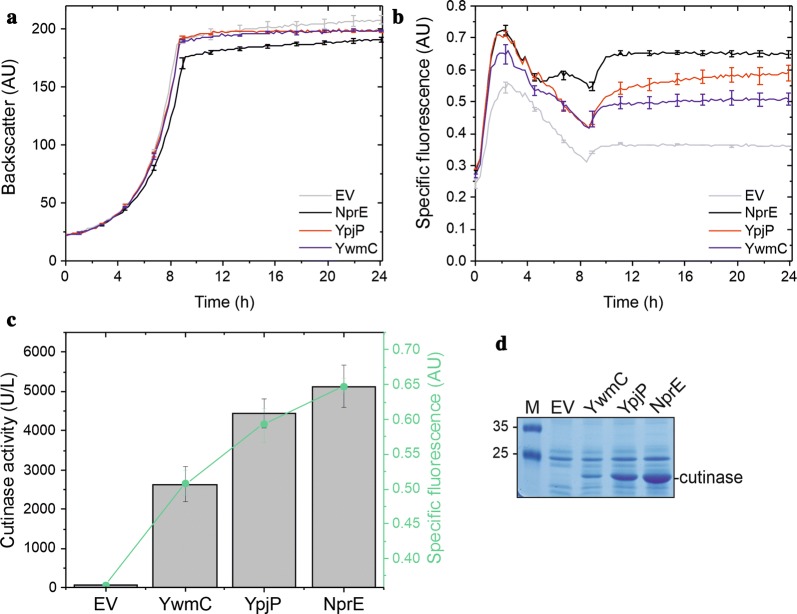
Fig. 4.
Cutinase secretion by C. glutamicum K9 using three different Sec signal peptides. Cells of C. glutamicum K9 possessing the pEKEx2 empty vector (EV), pEKEx2-NprE-cutinase (NprE), pEKEx2-YpjP-cutinase (YpjP), or pEKEx2-YwmC-cutinase (YwmC) were inoculated to an OD600 of 0.5 in 750 µl CGXII medium in 48-well FlowerPlates and subsequently cultivated in a BioLector system for 24 h at 30 °C, 1200 rpm and constant 85% relative humidity. After 4 h of cultivation, IPTG was added (250 µM final concentration). a Growth of the respective cultures was monitored as backscattered light in 15 min intervals starting at the beginning of the cultivation. The growth curves show one representative experiment of three independent biological replicates. Standard deviations are given for selected time points. b Specific fluorescence of the respective cultures during the BioLector cultivation. Also here, one representative experiment of three independent biological replicates is shown and the standard deviations are given for selected time points. c Cutinase activity in the supernatant (gray bars) and specific fluorescence values (green dots) after 24 h of cultivation. d After 24 h of growth, samples of the culture supernatant corresponding to an equal number of the respective C. glutamicum K9 cells were analyzed by SDS-PAGE and the proteins were visualized by Coomassie Brilliant Blue staining. M, molecular weight protein markers in kDa. The position of the secreted cutinase protein is indicated. AU arbitrary units

