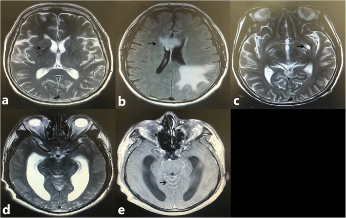
Fig. 1.
Neuroimaging characters of patients with cryptococcal meningitis. a T2-W image shows multiple dilated Virchow-Robin spaces (black arrow) in basal ganglia; b Abnormality (black arrow) on FLAIR image within the occipital lobe and corpus callosum; c T2-W image shows a gelatinous pseudocyst (black arrow) in basal ganglia; d hydrocephalus on T2-W image; e Contrast-enhanced image shows meningeal enhancement (black arrow) in cerebellum

