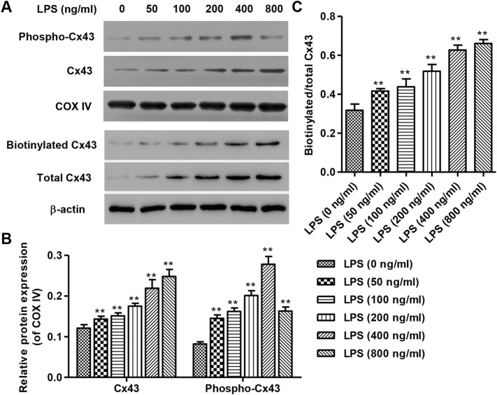
Fig. 1.
Expression of Cx43 and phospho-Cx43 after 24 h LPS stimulation. After continuous exposure to LPS for 24 h, protein levels of Cx43 in mitochondria and plasma membrane in HUVECs were detected by Western Blot (a), which showed concentration-dependent effects from 0 to 800 ng/mL, with 800 ng/mL LPS stimulation achieving the strongest expression of Cx43 (b, c). Biotinylated Cx43 is a representation of plasma membrane Cx43. Expression of mitochondrial phospho-Cx43 was detected by Western Blot (a), which likewise showed concentration-dependence effects from 0 to 400 ng/mL, with 400 ng/mL LPS stimulation achieving the strongest expression of phospho-Cx43 (b). COXIV and β-actin were used as loading control. Experiments were performed in 3 biological replicates and data are presented as mean ± SD. **P < 0.01 compared with 0 ng/ml LPS

