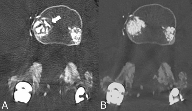
Fig 5.

A, Axial CT image with MAR applied demonstrating an irregular fragmented appearance of bone cement with indistinct margins (white arrow). B, Non-MAR counterpart image with normal appearance of bone cement with accurate representation of vertebral filling.
