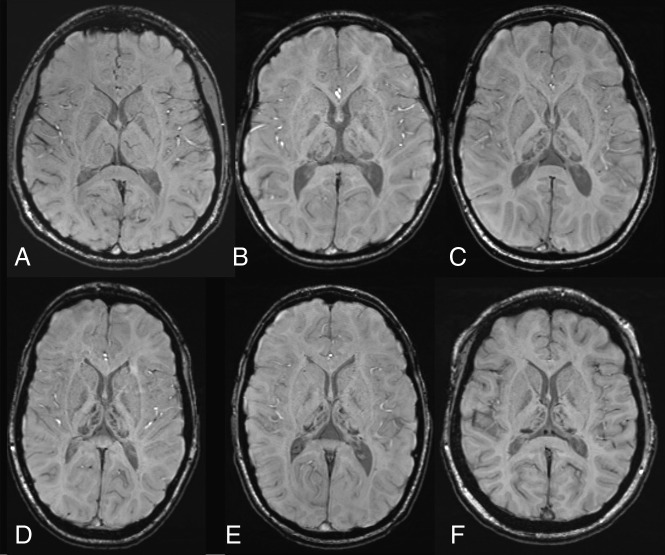
Fig 1.
Susceptibility-weighted axial images at the level of basal ganglia and thalami. A, A healthy control, an 11-year-old girl with normal findings. B–F, Five patients with AGU at different ages (7.9, 9.5, 11.4, 13.8, and 14.2 years) show a strikingly similar distribution of hypointensity in deep gray matter structures, with a maximum-intensity decrease in the pulvinar nuclei. Patchy decrease is also noted in the medial and anterior thalamic nuclei. Some signal intensity decrease is also seen in the globi pallidi.

