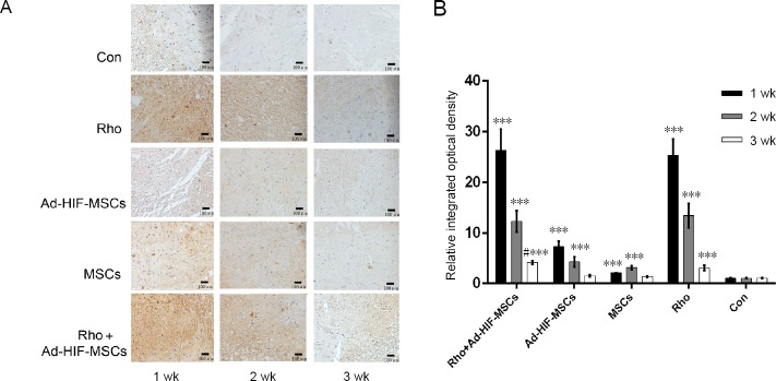
Figure 1.
Immunohistochemical staining of spinal cord tissue with an antibody against HIF-1 in each group at 1, 2, and 3 weeks after acute SCI.
(A) Immunohistochemical staining of spinal cord tissue (original magnification, 200×), scale bars: 100 μm. HIF-1 positive cells are dark brown. (B) Relative integrated optical density. ***P < 0.001, vs. control group; #P < 0.05, vs. Rho group. Data are expressed as the mean ± SD (n = 3; two-way repeated measures analysis of variance followed by Tukey’s post hoc test). Ad: Adenovirus; Con: control; HIF: hypoxia inducible factor; MSC: mesenchymal stem cell; Rho: rhodioloside.

