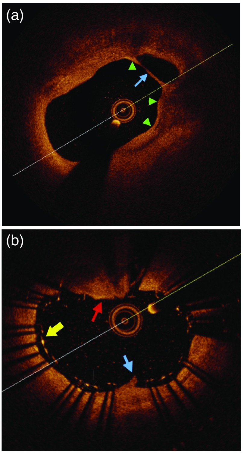
Fig. 1.
OCT imaging of carotid plaque before and after stenting. (a) Example of thin fibrous cap (green arrows) over a necrotic core (blue arrow) before stenting. These are both concerning plaque features increasing the future risk of stroke. (b) Poststenting example of stent-strut malapposition (yellow arrow), plaque prolapse through the stent (blue arrow), and thrombus over the stent (red arrow). These features are high-risk for stoke development.

