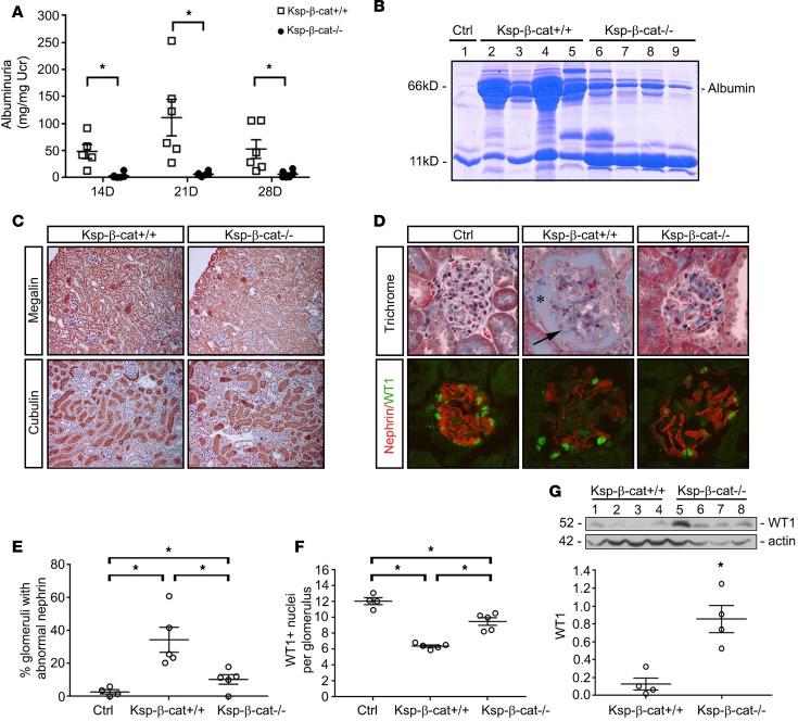
Figure 1. Tubule-specific β-catenin–knockout mice are protected from glomerular injury.
The β-catenin–floxed mice (Ksp-β-cat+/+) and tubule-specific β-catenin–knockout (Ksp-β-cat–/–) mice were subjected to continuous angiotensin II (Ang II) infusions (1.5 mg/kg/d, osmotic minipump). (A) Urinary albumin excretion was measured in the Ksp-β-cat+/+ and Ksp-β-cat–/– mice (n = 6) at 14, 21, and 28 days after Ang II infusion. *P < 0.05 compared with Ksp-β-cat+/+ at same time point, 1-way ANOVA. (B) Gel electrophoresis of urine samples shows the composition of the protein excreted in the urine. Albumin is indicated. Urine from an untreated control mouse (Ctrl, lane 1) was included for reference. (C) Levels of megalin and cubilin were not different between Ksp-β-cat+/+ and Ksp-β-cat–/– mice after Ang II infusion (original magnification, ×20). (D) Evaluation of glomerular histology reveals increased glomerular damage in Ksp-β-cat+/+ compared with Ksp-β-cat–/– mice. Note the high levels of protein in Bowman’s space in the Ksp-β-cat+/+ mice (asterisk), accompanied by marked glomerulosclerosis (arrow). In the Ksp-β-cat–/– mice, overall glomerular injury, including glomerulosclerosis, was minimal. A control (Ctrl, untreated mouse) glomerulus is provided for reference. Immunofluorescence revealed substantial disruption in nephrin and fewer WT1-positive podocytes in the Ksp-β-cat+/+ compared Ksp-β-cat–/– mice (original magnification, ×40). (E) Quantitation of the number of glomeruli with abnormal nephrin staining and (F) WT1-positive nuclei per glomeruli (n = 5, *P < 0.05, 1-way ANOVA). (G) Immunoblot for WT1 showing depleted levels in the Ksp-β-cat+/+ mice, compared with Ksp-β-cat–/– mice. *P < 0.05, t test.

