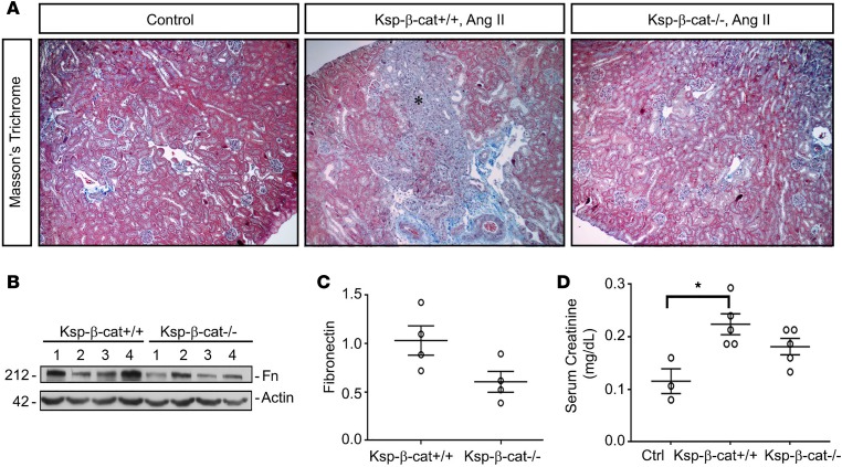
Figure 3. Ablation of β-catenin in renal tubules reduces interstitial fibrosis induced by Ang II infusion.
(A) Masson’s trichrome staining showing increased fibrosis in Ksp-β-cat+/+ mice after Ang II infusion (asterisk), compared with Ksp-β-cat–/– mice (original magnification, ×20). (B and C) Western blots show a trend toward increased levels of fibronectin in the control mice, compared with Ksp-β-cat–/– mice (n = 4). P = 0.06, t test. (D) Measurement of serum creatinine shows that the only significant difference is observed between untreated control mice (Ctrl) and Ksp-β-cat+/+ mice, suggesting moderation of injury in the Ksp-β-cat–/– mice (n = 5). *P < 0.05, 1-way ANOVA.

