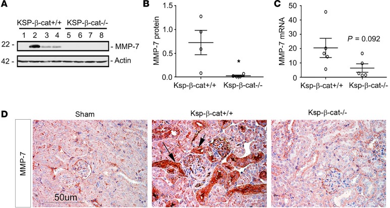
Figure 4. Renal expression of MMP-7 is reduced in Ksp-β-cat–/– mice after Ang II infusion.
(A and B) Western blot analyses for MMP-7 revealed significant reduction in the Ksp-β-cat–/– mice after Ang II infusion, compared with Ksp-β-cat+/+ mice (n = 4). Western blot (A) and quantitation after densitometry (B) are shown. *P < 0.05, t test. (C) qRT-PCR shows a trend to higher expression of MMP-7 mRNA in the Ksp-β-cat+/+ mice as well (n = 5, t test). P = 0.092, 2-tailed; P = 0.046, 1-tailed. (D) Immunohistochemical staining for MMP-7 shows negligible staining in control mice, with dramatic upregulation in Ang II–treated Ksp-β-cat+/+ mice (arrows). A reduced MMP-7 staining was noticed in Ksp-β-cat–/– mice. Scale bar: 50 μM.

