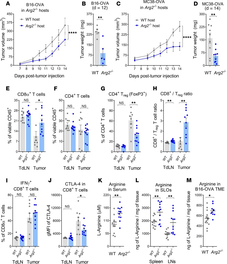
Figure 1. Deletion of Arg2 reduces tumor growth and increases arginine availability.
(A–D) Analysis of tumor growth (A) for B16-OVA (n = 10) and (C) for MC38-OVA (n = 13) and tumor weight at (B) day 12 or (D) day 14 tumors in WT or Arg2–/– hosts. (E–I) T cell frequencies in tumor-draining lymph nodes (TdLN) and tumors in 9-day MC38-OVA tumor-bearing WT and Arg2–/– mice: (E) CD8+ T cells; (F) CD4+ T cells; (G) FoxP3+ Tregs; (H) CD8+/FoxP3+ T cell ratio; (I) IFN-γ+CD8+ T cells. (J) Analysis of CTLA-4 expression by CD8+ T cells. (K–M) HPLC-MS quantification of arginine in (K) serum, (L) secondary lymphoid organs (SLO), and (M) B16-OVA tumors 12 days after tumor implantation in WT or Arg2–/– mice. Results were pooled from 2 or 3 independent experiments (A, C, E–M) or are representative of 3 independent experiments (B and D). Data is represented as mean ± SEM throughout. *P < 0.05, **P < 0.01, and ****P < 0.0001 (A and C: 2-way ANOVA) (B, D–M: 2-tailed Student’s t test).

