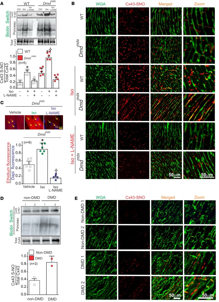
Figure 3. Iso increases S-nitrosylated levels of Cx43 at the lateral side of Dmdmdx cardiomyocytes.
(A) Top and middle gels were loaded with S-nitrosylated proteins pulled down from heart samples using the biotin switch assay. Top gel was, then, blotted against Cx43, and the middle gel is the corresponding Ponceau staining. Lower blot was loaded using total cardiac proteins and blotted against Cx43. The bottom graph is the quantification for 5 independent blots using the ratio for S-nitrosylated Cx43/Ponceau. The number in parentheses indicates the n value. Comparisons between groups were made using 2-way ANOVA plus Tukey’s post hoc test. *P < 0.05 vs. WT control, and †P < 0.05 vs. WT Iso. (B) Analysis performed by PLA of the interaction between Cx43 and S-nitrosylation. Plasma membrane stained with wheat germ agglutinin (WGA) and S-nitrosylated Cx43 (Cx43-SNO) are shown in green and red, respectively. Representative images of n = 5 per group. (C) Assessment of Cx43 hemichannel activity in isolated Dmdmdx hearts perfused with buffer containing 5 μM ethidium bromide after or without treatment with Iso. Arrows show nuclei. The number in parentheses indicates the n value. Comparisons between groups were made using 2-way ANOVA test plus Tukey’s post hoc test. *P < 0.05 vs. vehicle WT; †P < 0.05 vs. vehicle Dmdmdx. (D) Top and middle gels were loaded with S-nitrosylated proteins pulled down from human heart samples using the biotin switch assay. Top gel was, then, blot against Cx43 and the middle gel is the corresponding ponceau staining. Lower blot was load using total cardiac proteins and blot against Cx43. (E) Analysis performed by PLA of the interaction between Cx43 and S-nitrosylation in human samples. Note that Cx43 is S-nitrosylated at the lateral side of DMD human samples compare to non-DMD.

