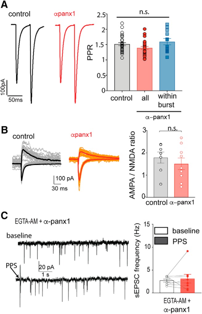
Figure 7.

The block of postsynaptic Panx1-induced increase in sEPSCs does not alter evoked EPSCs, AMPAR/NMDAR ratios but requires increased Ca2+. A, PPR is not altered by blocking postsynaptic Panx1. Average (n = 15 pairs) PPRs (at 0.05 Hz, 1 ms duration, 50 ms interstim interval) are shown under control (black) and with α-panx1 in the pipette (red). Right, The population response of the PPR. In the presence of α-panx1 in the pipette (n = 31), the PPR over the 5 min recordings was unchanged compared with the control (n = 28). When the increased sEPSC frequency overlapped with the evoked responses (blue bar; n = 12), the PPR was still unchanged. B, AMPAR/NMDAR ratio is not altered by block of postsynaptic Panx1. Families from both control (normal internal solution; n = 7) and with α-panx1 (n = 9) in the pipette (red) are shown. Downward traces represent the AMPAR component collected at Vm = −70 mV in 1.2 mm Mg2+. Upward traces represent the NMDAR component collected at Vm = 40 mV in 0 Mg2+ and 10 μm DNQX. Right, Calculated ratios. n.s., Not significant. C, Incubation of hippocampal slices with EGTA-AM prevented the increased sEPSC frequency arising from Schaffer collateral stimulation when α-panx1 was in the pipette. Example 10 s traces are shown. Arrow indicates PPS. Right, The plot illustrates a failure of the mean frequency of sEPSCs to increase, suggesting that bursting is Ca2+-dependent.
