Figure 5.
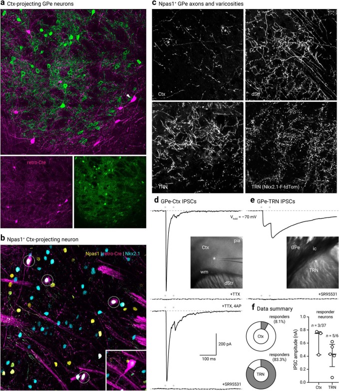
Cortex-projecting neuron properties. a, LV retrograde labeled (magenta) GPe neurons with PV immunostaining (green). Note that cortical projecting GPe neurons are not PV+. Arrowhead indicates a LV-labeled neuron with a large cell body characteristic of cholinergic neurons. b, Confocal micrograph showing the coexpression (dotted circles) of Npas1 (yellow) and Nkx2.1 (blue) in cortex-projecting GPe neurons (magenta). Inset: an example of a neuron (shown at the same magnification) that has a large cell body and low Nkx2.1 expression, features of cholinergic neurons within the confines of the GPe. c, High-magnification confocal micrographs of axons in the Ctx, dStr, and TRN with injection of a CreOn-ChR2 AAV into the GPe of a Npas1-Cre-tdTom mouse. Asterisks in the top left denote putative terminals. Bottom right, high density of synaptic boutons in the TRN of Nkx2.1-F-tdTom mice. d, Voltage-clamp recordings of the Npas1+ input in a cortical neuron within layers 5 and 6. The recorded neuron was held at -70 mV with a high Cl− internal; IPSCs (IPSCs) were evoked from 20 Hz paired-pulse blue light stimulation (indicated by gray circles). Note the fast and depressing responses evoked. Inset: location of the recorded neuron (asterisk) in the Ctx is shown. IPSCs were attenuated with extracortical stimulation (data not shown) and abolished with tetrodotoxin (TTX, 1 μm). Application of 4-aminopyridine (4-AP, 100 μm) in the presence of TTX restored the response with intracortical stimulation. IPSCs were completely blocked with SR95531 (10 μm). e, Voltage-clamp recording of a TRN neuron with identical experimental setup shown in d, Note the facilitating responses evoked. Inset: location of the recorded neuron (asterisk) is shown. Responses were sensitive to the application of SR95531 (10 μm). f, Left, Pie charts summarizing the percentages of responders in Ctx and TRN. Right, Medians and interquartile ranges of IPSC amplitudes are represented in a graphical format. dStr, dorsal striatum; Ctx, cortex; TRN, thalamic reticular nucleus; ic, internal capsule; wm, white matter.

