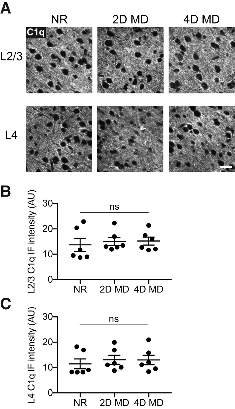
Figure 7.

C1q levels in V1b do not change with monocular deprivation. A, Representative images of C1q immunohistochemistry in L2/3 (top) and L4 (bottom) of V1b, from P32 mice that were NR, or from animals that had undergone 2 d (2D MD) or 4 d (4D MD) of MD starting at P28. Images are from the hemisphere contralateral to the deprived eye. Scale bar, 20 μm. B, Quantification of C1q immunofluorescence intensities in L2/3 of V1b in NR, 2D MD, and 4D MD animals. The conditions are not statistically different from one another (one-way ANOVA, F(2,15) = 0.1803, p = 0.8368, n = 6 animals/condition). C, Quantification of C1q immunofluorescence intensities in L4 of V1b in NR, 2D MD, and 4D MD animals. There is no statistically significant difference in immunofluorescence intensities between the conditions (one-way ANOVA, F(2,15) = 0.2338, p = 0.7943, n = 6 animals/condition). All error bars represent SEM. ns, not significant.
