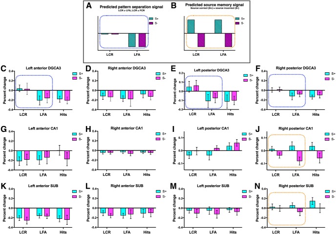
Figure 3.
Dissociated pattern separation and source memory signals in hippocampal subfields and MTL subregions. A, Schematic of the predicted pattern separation signal (blue dots), where activity for LCRs is on par with foil correct rejections (FCR, baseline) and LFAs show repetition suppression (LCR > LFA). B, Schematic of the predicted source memory signal (orange dots), where activity for correct source judgments is greater than for incorrect source judgments. Activity in the left anterior DG/CA3 (C) and posterior DG/CA3 (E,F) was greater for correct rejections, consistent with a pattern separation signal. Activity in the right posterior CA1 (J) was greater for correct source memory judgments, consistent with a source memory signal. N, Marginal effect of source in the right posterior SUB (p = 0.065). Activity in the left posterior CA1 (I) showed a response profile that was not consistent with predicted pattern separation or source memory signals. When target hits were included in the analysis, there was an effect of item memory and source in this region, with greater activity for target hits than for LCR and LFA. Dotted rectangle represents conditions that were included in the two-way ANOVA. Activity in all other hippocampal ROIs is also presented (D, G, H, K–M). Foil correct rejections were used as baseline. Error bars indicate SEM. SUB, Subiculum.

