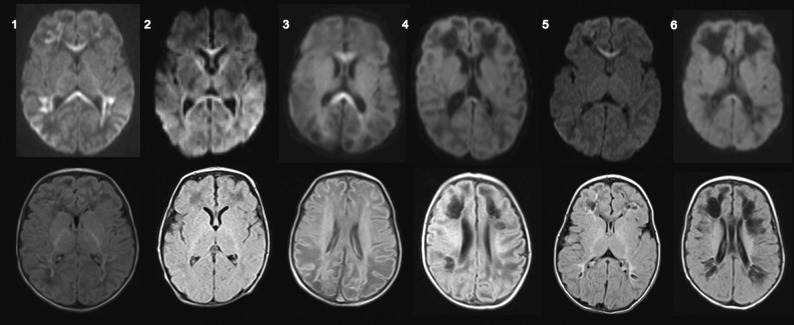
FIGURE.
Brain MR imaging of 6 infants with confirmed perinatal CHIKV infection. Axial diffusion-weighted imaging (upper row) and axial FLAIR (lower row) of patients 1–6. Patients who underwent MR imaging before 14 days of symptom onset presented with restricted diffusion in the corpus callosum and/or subcortical white matter with a perivascular distribution, whereas patients who underwent MR imaging after 14 days of symptom onset presented with subcortical cystic lesions, also with a perivascular distribution with discrete restricted diffusion in the corpus callosum.

