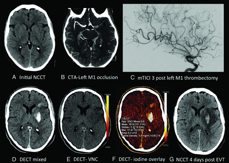
Fig 2.
A 59-year-old man with a preprocedural ASPECTS of 8 (A), left M1 occlusion (B), and modified TICI 3 reperfusion (C). Postthrombectomy DECT demonstrates parenchymal hyperdensity in the left basal ganglia and frontal lobe (D), without hemorrhage on the virtual noncontrast DECT (E) and consistent with contrast staining on the iodine overlay map (F), with maximum iodine concentration measuring 3.4 mg/mL and 428.2% relative to the SSS. NCCT performed 4 days postthrombectomy (G) demonstrates evolving left MCA infarction with development of grade 3 ICH involving the left lentiform nucleus.

