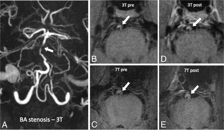
Fig 3.
A 73-year-old man with a posterior circulation stroke, an occluded left vertebral artery, and stenosis of the basilar artery. TOF angiography at 3T of a patient with basilar artery stenosis (white arrow, A). Pre- and postcontrast vessel wall imaging at 3T (B and C) and 7T (D and E) in a patient with basilar artery atherosclerotic plaque (white arrows indicate areas of stenosis).

